

microscope and accessories (tweezers, microscope slide, cover slip, dissecting needles, pipette), a red onion, water, saline solution
First, the students should prepare a red onion for the observation with the microscope (see worksheet 7). The teacher gives the following instruction: “Prepare a red onion as described in worksheet 7 and look at it with the microscope. Then consider a second onion preparation that lies in salt solution instead of water on the microscope slide. What can you see?“
During this task, the students can observe the composition of life (and living creatures) from cells and develop an understanding of the impact of salt water.
Background information: Plasmolysis, cell structure, microscope structure (see worksheet 7)
Observation 1: The epidermis of the red onion consists of cells, too. There is a red area in each cell.
Explanation: The red area, which sometimes seems to fill in the whole cell, is a kind of storage room for nutrient-containing cell sap.
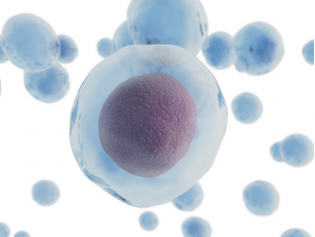
Pic 91: Onion cell 2
Observation 2: The red area of the onion cells contracts more and more in the salt solution. It is partly possible to see that a thin membrane detaches from the cell wall.
Explanation: The higher salt concentration outside the cells detracts liquid from the cells.